Among the many varieties of microscopes, optical and electronic are considered the most popular. The first option refers to traditional optical equipment. Such devices make it possible to distinguish between particles up to 0.2 µm. Their magnifying ability can reach 2000 times.
Electron microscopes are more powerful. The technology implemented in them allows you to achieve an increase of 200,000 times. These devices use an electron beam. Special magnetic lenses are used to obtain an image. This technique is more relevant for most consumers. Electron microscopes are raster (scanning) and translucent. Some models combine the properties of these two types of equipment.
The scope of this technology is very wide. Electron microscopy is used to study biofilms of microorganisms and other materials. This research method is in demand in science, industry, forensic examination and many other areas.
Focused beam JEOL (Japan)
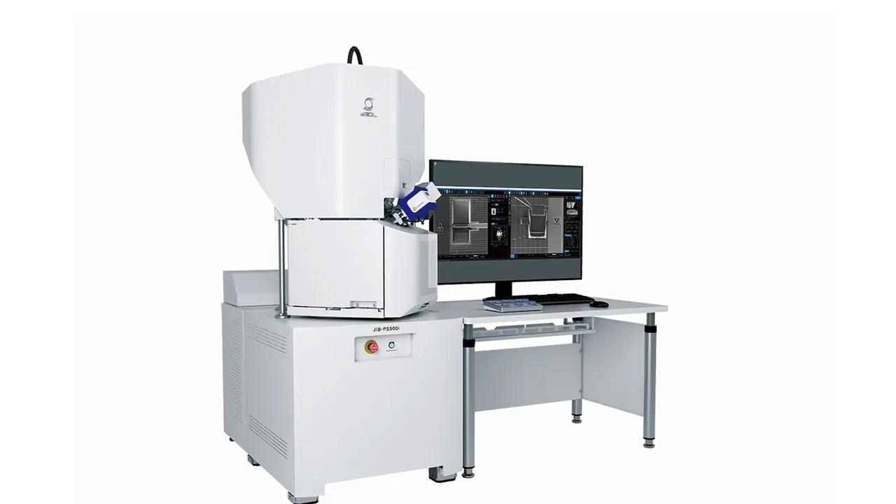
JEOL has developed a new Focused Ion Beam (FIB) solution for preparation of specimens prior to observation in the Transmission Electron Microscope (TEM). The new JIB-PS500i is a multipurpose FIB-SEM that delivers the synergy of fast sample preparation, SEM imaging and EDS analysis in a single instrument.
High-Quality Fast TEM Sample Preparation
The new FIB sample stage offers fast transitioning between processing and imaging, allowing for real-time feedback of specimen quality. With the ability to prepare samples thinner than 30nm, the FIB-SEM produces a sample suitable for superior atomic resolution imaging and analysis with STEM (Scanning Transmission Electron Microscope). A retractable STEM detector enables easy acquisition of bright field and dark field images during processing to precisely evaluate preparation of the TEM sample. The operator can easily prepare TEM specimens using the STEMPLING2 automatic TEM specimen preparation system, which allows unattended preparation of multiple samples.
A specially designed double-tilt sample holder, TEM-Linkage, enables seamless transfer from the FIB-SEM directly to the TEM.
New Large Chamber/Stage for Ultimate Sample Preparation Flexibility
A key advantage of the JIB-PS500i FIB is the large specimen chamber with an easy-access door. This design supports an efficient workflow and flexibility for a variety of samples and processes. The 5-axis full-eucentric large motor stage is designed to transport both large and multiple samples in the XY direction, and at a wide stage tilt and rotation range.
New FIB Column with higher current and superior performance at low kV
A new high current (up to 100 nA) FIB column is especially effective for large-area processing and analysis, which is ideal for semiconductor samples. The new FIB has high performance fine milling capabilities essential for quality lamella preparation imaging, EDS analysis, and 3D microscopy. The new JIB-PS500i has superior performance in the low kV range, as low as 0.5kV, essential for beam sensitive materials.
JEOL Transmission Electron Microscopes
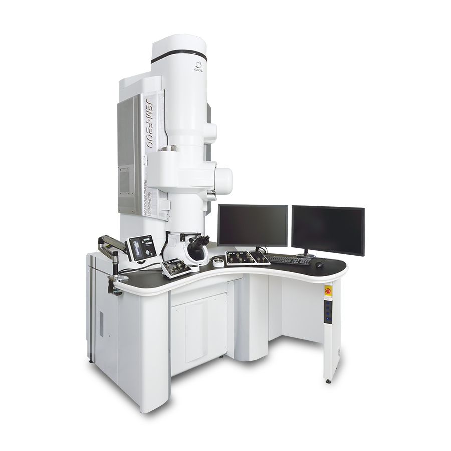
For in-depth research work and obtaining high-resolution images in the field of semiconductor industry, nanotechnology, materials science, it is necessary to use a device for an ultrathin sample by passing an electron beam through it. The transmission electron microscope is excellent for solving these problems. Such devices are used in the work of many research institutes, enterprises, biomedical laboratories, etc.
The device is aimed at solving analytical problems. The presented models obtain filtered images and electronic spectra without significant energy losses. The equipment receives the necessary data without distortion due to the use of a reliable optical-electronic system.
Optical microscopes Nikon (Japan)
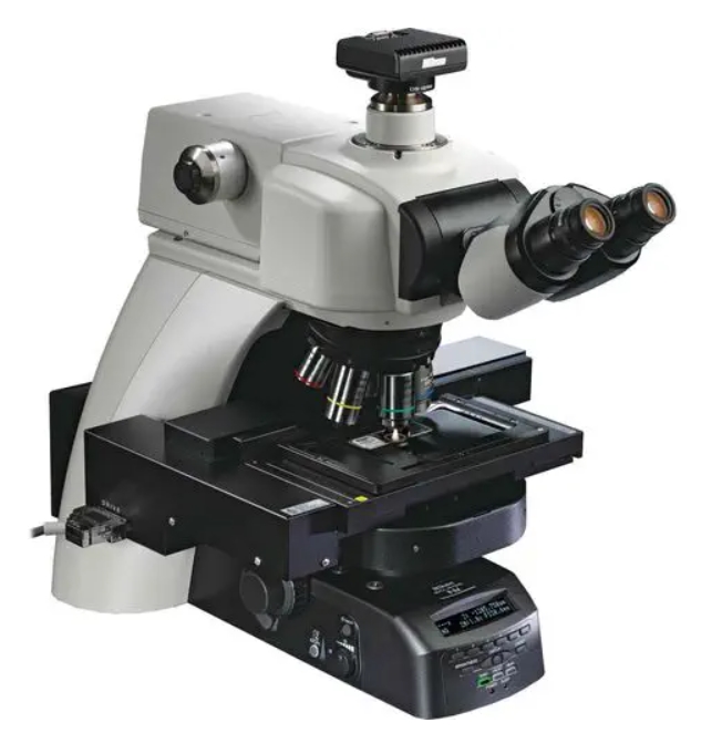
Fully motorized and advanced research upright microscope offering automated acquisition capabilities of multi-dimensional images Eclipse Ni-E, Nikon’s top-of-the-line motorized upright microscope, was developed to meet the increasing demands for sophistication and automation of research. Eclipse Ni-E boasts flexible system expandability and supports a broad range of advanced research applications.
With Nikon’s proprietary stratum structure, exchangeable focusing mechanisms, from focusing stage to focusing nosepiece, a diverse array of motorized accessories, and automatically changing observation conditions, Ni-E has expanded application possibilities to provide the perfect solution for all advanced bioscience and medical research.
Key Features Stratum structure improves system expandability Nikon’s proprietary stratum structure enables simultaneous mounting of two optical paths on one microscope to support various applications.
This structure allows double layer mounting of an epi-fluorescence illuminator and a barrier filter wheel, or a photoactivation unit and a back port unit, developed for the first time for upright microscopes.
Focusing mechanisms can be modified for the application The microscope can be re-configured by switching the focusing stage and focusing nosepiece, enabling a fixed-stage configuration with motorized focusing to meet the demands of experiments such as in vivodeep imaging utilizing multiphoton confocal microscopy.
Scanning electron microscopes
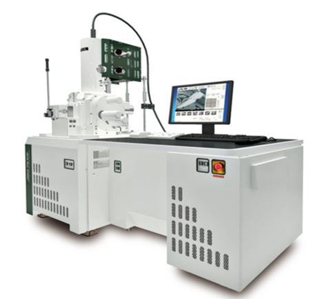
Laboratories and industrial enterprises need to conduct various studies, where it is necessary to obtain high-resolution images of the surface of an object, as well as information on the composition, structure, and some other properties. To perform this task, you need to use an electron microscope class device. A scanning electron (scanning) microscope is suitable for this. You can buy this equipment from us on favorable terms.
Surface analyzers JEOL (Japan)
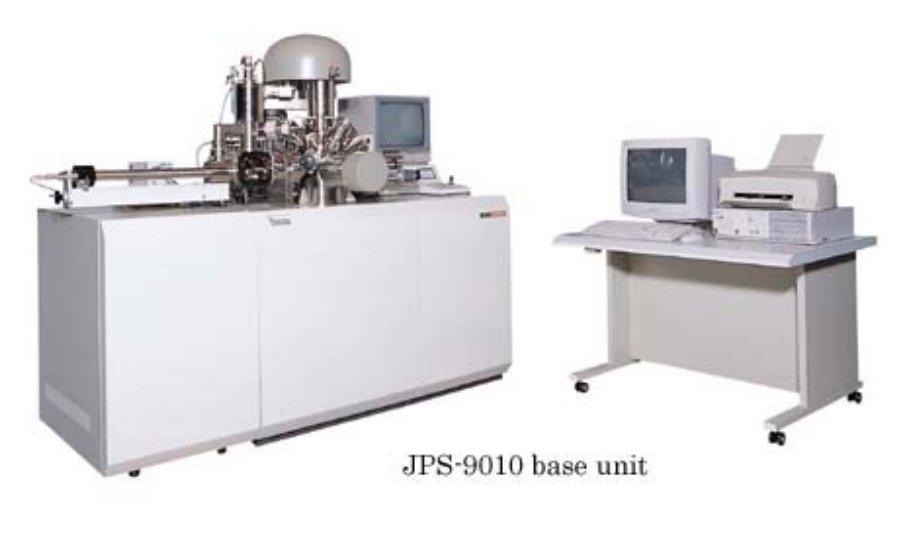
JPS-9010 Series - Features
The JPS-9010 series offers a variety of accessories to meet a wide range of applications including high resolution measurement, angle resolved XPS analysis, and depth profiling.
- High accuracy energy analyzer
- X-ray source to minimize damage to samples
- Compact X-ray monochromator: Neutralizing gun incorporated
- Ultra high clean vacuum system; easy baking
- Versatile auto analysis to support routine analysis
- Large stage to accommodate a 3.5” hard disk
- Easy to use software: Windows®XP compatible
- High speed peak separation software
- High speed ion gun: Rapid depth profiling at low acceleration voltage and high current
